Clinical History
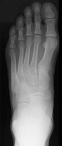
A 22 year old woman presents with a two week history of pain in her left forefoot. She is a fit woman who normally goes to the gym twice a week and has just begun training for the City to Surf in August this year. She has been running about 5 km twice a week for the past month and cannot remember any traumatic event to cause her foot to be painful. At rest, the pain is not particularly severe but worsens quickly during exercise. Plain radiographs of her
left foot are normal and a bone scan was performed.
Scan Findings
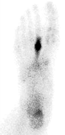
There is a recent stress fracture involving the shaft of the second metatarsal on the left, corresponding to the site of her pain.
Discussion
Stress fractures are among the most common sports injuries and should be suspected in any patient with localised forefoot pain. This is particularly the case in patients who have recently started an exercise program or increased the
intensity of the exercise in preparation for an event.
Metatarsal stress fractures account for approximately 25% of stress fractures and most often involve the distal portions of the second and third metatarsals. Plain radiographs are relatively insensitive in detecting stress fractures at the time
of injury and can only pick up about 50% of fractures in the convalescent phase when performed at 10 weeks after the onset of pain. Bone scans are very sensitive to the detection of stress fractures and a normal bone scan reliably excludes
a stress fracture. Metatarsal stress fractures involving the second and third metatarsal usually heal well when treated with immobilization in an othopaedic boot for four to eight weeks. Fractures of the fifth metatarsal can be more problematic as these fractures are prone to non union.
Approximately 60 percent of patients with a stress fracture have had a previous stress fracture and there are studies suggesting that gradually increasing in the intensity of exercise, pre exercise stretching and the use of shock absorbing insoles may prevent future fracture.
Conclusion
Stress fractures generally occur as a result of repetitive use that exceeds the intrinsic ability of the bone to repair itself. In an athlete complaining of localised pain in the foot, a stress fracture is the most likely diagnosis and can be easily confirmed or excluded by a bone scan.
Reference: Sanderlin BW, et al. Common stress fractures. Am Fam Physician 2003; 68:1527-32
Case Study submitted by
Patrick Butler
Department of Nuclear Medicine,
(PDF DOWNLOAD)
Figure 1. Plain radiograph of left foot with no evidence of fracture. Oblique views also failed to demonstrate any abnormality.
Figure 1. Plantar view of a bone scan showing marked focal accumulation of tracer in the mid shaft of the second left metatarsal with extension of tracer into the shaft of the
metatarsal. In addition, there is mildly increased uptake in the calcaneus. The patient was not tender at this site and this abnormality is explained by the altered weight bearing caused by walking with a painful forefoot.