Clinical History
A 55 year old lady complains of left wrist pain following excessive gardening. The symptoms are located at the base of the thumb where there is boggy tenderness and are exacerbated by thumb movement. She is left handed and has had long standing left thumb base pain, however this recent pain is different. A radiograph shows left 1st carpometacarpal (CMC) joint degenerative arthritis. It is unclear whether the symptoms relate to the degenerative arthritis or another cause and a bone scan is arranged.
Scan Findings
A three phase bone scan was performed. In a three phase bone scan, the first phase (blood flow phase - not shown here) is acquired immediately following tracer injection and shows hyperaemia. The next phase (blood pool phase) demonstrates inflammation. Both these phases give information regarding the soft tissues, whereas the final phase (delayed phase) mainly demonstrates bone activity.
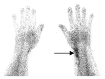
In this patient, there is a line of increased blood pool
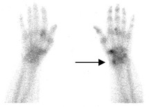
activity (Fig.1) in the radial aspect of the left wrist typical for tendon inflammation. There is low grade uptake in the periosteal edge of the left distal radius in the delayed images (Fig.2). This is not a bone abnormality per se but represents reactive uptake in the bone underlying the inflamed tendons.
Discussion
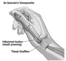
This is a case of radial tenosynovitis (de Quervain’s) due to inflammation of the thumb tendons - abductor pollicis longus and extensor pollicis brevis. Repetitive use of the thumb can be a contributing factor and it commonly occurs following excessive activity. Although symptoms are thought to result from inflammation, pathology often shows a chronic degenerative process. Bone scans are useful for diagnosis where the clinical scenario is unclear and are particularly helpful in excluding other causes such as fracture or arthritis. The increased blood pool seen in the early (blood pool) phase of the study compared with the delayed phase is typical for a predominant soft tissue process, in this case tendonitis and helps distinguish primary bone pathology from soft tissue pathology. The three phase bone scan is useful in other soft tissue abnormalities such as bursitis, synovitis, fascial inflammation, muscle injury and enthesopathy.
Conclusion
Many soft tissue abnormalities can be demonstrated in a three phase bone scan. In this case radial tenosynovitis is confirmed with no evidence for fracture or significant arthritis.
Case Study submitted by
Dr Scott Beuzeville
Department of Nuclear Medicine,
St. George Hospital.
Figure 1. Linearly increased blood pool activity indicating inflammation in the left radial wrist (arrowed).
Figure 2. Delayed images showing faint increased uptake in the periosteal edge of the left distal radius (arrowed) with low grade left 1st CMC joint degenerative arthritis just distal to this focus.
Figure 3. Diagram indicating the involved tendons in de Quervain’s tenosynovitis.